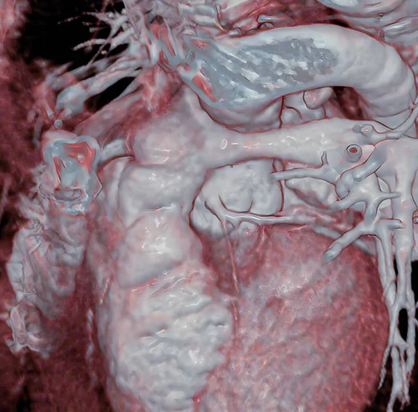
Heart Coronary Arteries Scan — Stockfoto

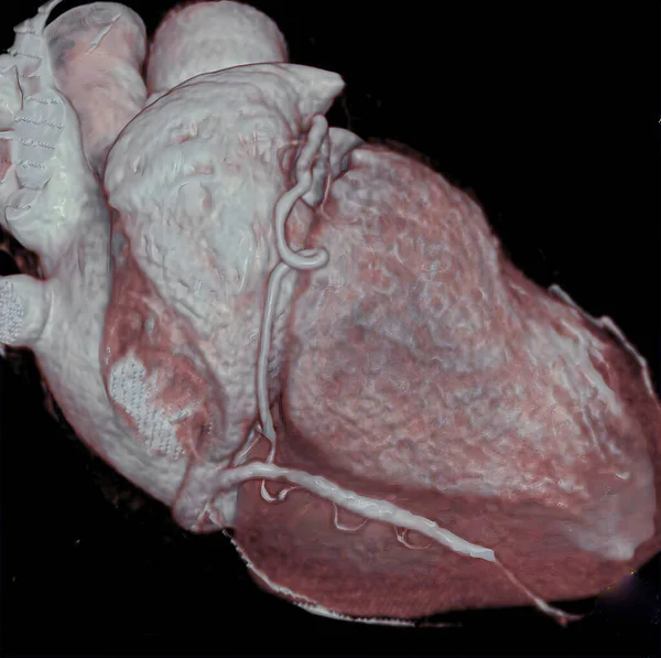
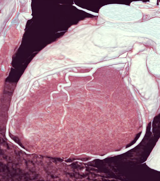
Heart and coronary arteries, 3D CT scan. — Stockbild

Heart and coronary arteries, 3D CT scan.

 — Foto av imagepointfr

Samma serie:

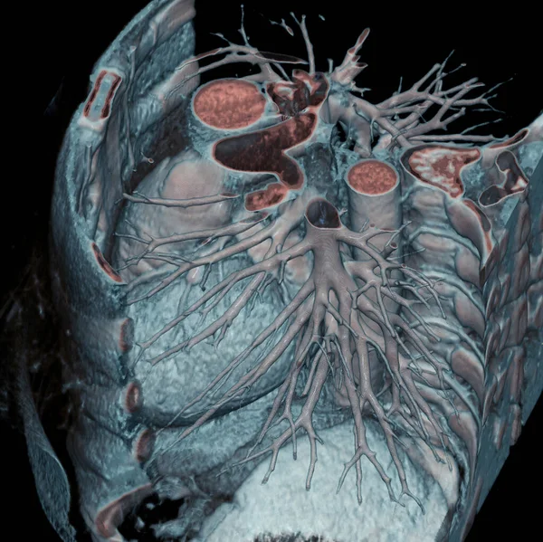
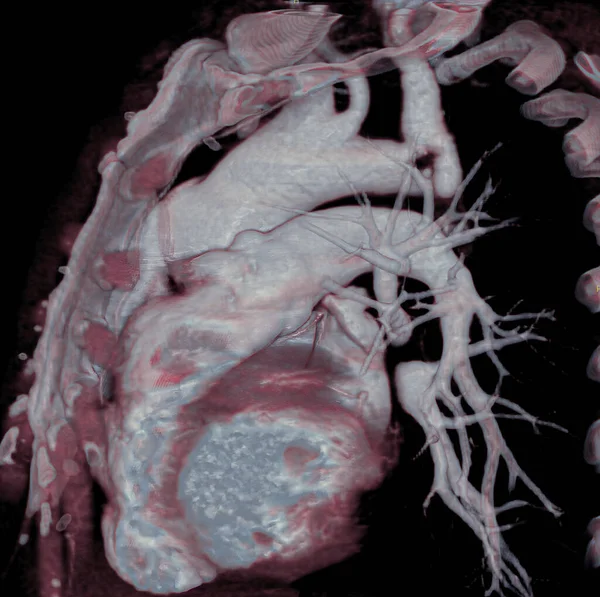
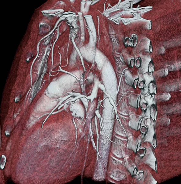
Thorax: Heart, aorta, and pulmonary artery are visible, 3D CT scan.
Thorax Heart Aorta Pulmonary Artery Visible Scan — Stockfoto
Anomalous left coronary artery, to the right of the aorta, passes in front of the pulmonary artery. This rare congenital anomaly is, nevertheless, not dangerous since the pulmonary artery does not restrict the coronary artery. 3D CT scan.
Anomalous Left Coronary Artery Right Aorta Passes Front Pulmonary Artery — Stockfoto
In patient born without pulmonary arteries, surgery reconstructs an almost-normal pulmonary artery tree by attaching native pulmonary, arterial branches to a tube placed in the right ventricle. 3D CT scan.
Patient Born Pulmonary Arteries Surgery Reconstructs Almost Normal Pulmonary Artery — Stockfoto
Heart and right coronary artery, 3D CT scan.
Heart Right Coronary Artery Scan — Stockfoto
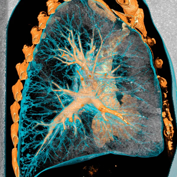
Pulmonary arteries and Mediastinum, heart, and aorta are visible, 3D CT scan.
Pulmonary Arteries Mediastinum Heart Aorta Visible Scan — Stockfoto
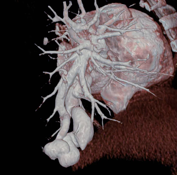
Abnormal vessel originating from the abdominal aorta and vascularizing the right lung. This anomaly, called pulmonary sequestration, can cause pulmonary hemorrhaging (hemoptysis). 3D CT scan.
Abnormal Vessel Originating Abdominal Aorta Vascularizing Right Lung Anomaly Called — Stockfoto
3D CT scan of normal coronary arteries.
Scan Normal Coronary Arteries — Stockfoto
3D CT scan of coronary bypasses.
Scan Coronary Bypasses — Stockfoto
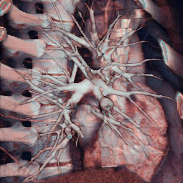
Stenosis of the pulmonary arteries. The 3D CT scan shows few pulmonary arteries. Some are occluded due to a pulmonary embolism.
Stenosis Pulmonary Arteries Scan Shows Few Pulmonary Arteries Some Occluded — Stockfoto
Pulmonary artery (PA) aneurysm. Pulmonary aneurysm is associated with pulmonary artery dilation, with a risk of intra-pulmonary hemorrhaging and rupture. 3D CT scan.
Pulmonary Artery Aneurysm Pulmonary Aneurysm Associated Pulmonary Artery Dilation Risk — Stockfoto
3D CT scan of coronary arteries and aorta of heart.
Scan Coronary Arteries Aorta Heart — Stockfoto
3D scanner of thoracic anatomy.
Scanner Thoracic Anatomy — Stockfoto
Pulmonary arteriovenous fistula is a congenital anomaly consisting of a direct communication between the pulmonary arterial and venous systems. Complications include intra-pulmonary hemorrhaging (hemoptysis). 3D CT scan.
Pulmonary Arteriovenous Fistula Congenital Anomaly Consisting Direct Communication Pulmonary Arterial — Stockfoto
Thorax: 3D CT scan.
Thorax Scan — Stockfoto
In the arterial switch operation (ASO), the aorta and the pulmonary artery are cut and reconnected to the \correct\ pumping chamber (ventricle). At birth the large vessels were reversed: the aorta originated in the right ventricle and the pulmonary a
Arterial Switch Operation Aso Aorta Pulmonary Artery Cut Reconnected Correct — Stockfoto
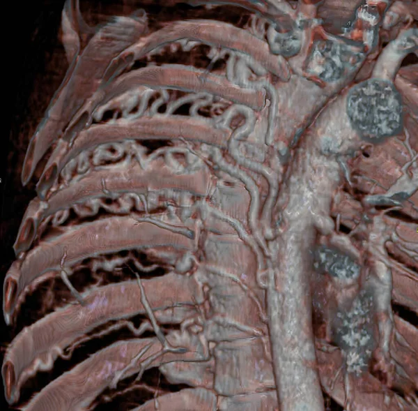
Large intercostal arteries, visible in a CT scan, developed to compensate pulmonary blood flow due to a congenital heart defect characterized by atresia of pulmonary arteries (small or absent pulmonary arteries). 3D CT scan.
Large Intercostal Arteries Visible Scan Developed Compensate Pulmonary Blood Flow — Stockfoto

Användningsinformation

Du kan använda det royaltyfria fotot "Heart and coronary arteries, 3D CT scan." för personliga och kommersiella ändamål enligt standardlicensen eller den utökade licensen. Standardlicensen omfattar de flesta användningsområden, inklusive reklam, design av användargränssnitt och produktförpackningar, och tillåter upp till 500 000 tryckta kopior. Den utökade licensen omfattar samtliga av standardlicensens användningsområden med obegränsade utskriftsrättigheter, och låter dig använda de nedladdade bilderna för handelsvaror, återförsäljning av produkter eller gratis distribution.

Vektorbilden kan skalas till valfri storlek. Du kan köpa och ladda ner den i hög upplösning upp till 4996x5003. Uppladdningsdatum: 11 aug. 2022